June 24, 2013

Innovative nanoparticle therapy to treat infected burn wounds

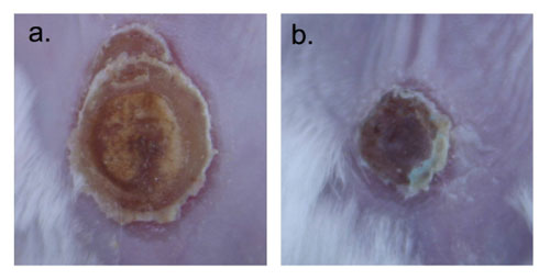
(Nanowerk Spotlight) Despite significant advances in the medical/surgical management of severe thermal injury, wound infection and subsequent sepsis persist as frequent causes of morbidity and mortality for burn victims not only due to the extensive compromise of the protective barrier against microbial invasion, but also as a result of growing pathogen resistance to our therapeutic options.Burn wounds are further complicated by injury induced suppression of the immune system, further facilitating localized colonization and systemic entry of microbes from both endogenous and exogenous origins. Candida yeast species represent a prevalent threat to these patients, many variants demonstrating resistance to first line systemic antifungal treatments as these medications are not directly toxic, but instead inhibit yeast growth through a single mechanism of action.Therefore, innovative therapies are urgently needed that overcome mechanisms of pathogen resistance, and are easily administered without concerning systemic side effects.Nanoparticles represent a practical platform through which both established drugs and novel agents can be encapsulated and delivered more effectively or via new routes to target and reach pathogen cells/machinery with greater success then their macromolecular counterparts.In a paper published in the June 14, 2013 online edition of Nanomedicine: Nanotechnology, Biology and Medicine ("Amphotericin B releasing nanoparticle topical treatment of Candida spp. in the setting of a burn wound") we demonstrated that encapsulating Amphotericin B, a intravenously administered potent fungicidal polyene macrolide, in nanoparticles increased its killing impact against numerous candida species, was more effective at preventing candidal biofilm formation, and cleared a mouse burn model infected with candida more effectively than solubilized amphotericin.LundImages were taken on day 9 after infection. a) is topically applied solubilized Amphotericin B and b) Amphotericin B nanoparticle applied. (Images: Dr. Friedman, Montefiore - Albert Einstein College of Medicine)Materials at the nanoscale have a greater likelihood of interacting with their desired target, here, the cell wall, and therefore the payloads have greater efficacy. Furthermore, the nanoparticles themselves can physically damage pathogen structures and prevent cell-to-cell communication and sharing of resources, offering synergists but different antifungal properties.Encapsulating amphotericin in nanoparticles not only made it more effective, but also allowed for it to be directly and topically applied to a wound – an option not currently available. Systemic delivery, while very effective, comes at a great cost as amphotericin has multiple well established side effects and toxicities associated with its use.Our team here at Einstein represents a broad range of expertise calling from multiple departments including medicine (infectious diseases, dermatology), physiology and biophysics, and immunology and microbiology, highlighting the multidisciplinary nature of nanotechnology research. Other authors included David Sanchez; David Schairer; Chaim Tuckman-Vernon; Jason Chouake; Allison Kutner; Joy Makdisi; Joel Friedman; and Joshua d Nosanchuk.By Dr. Adam Friedman, Assistant Professor of Medicine (Dermatology)/ Physiology and Biophysics, and Director of Dermatologic Research at Montefiore Medical Center - Albert Einstein College of Medicine.


Read more: http://www.nanowerk.com/spotlight/spotid=30997.php#ixzz2X9QjwHzd

April 12, 2013

Regenerative Heart Medicine Could Get Boost With Nanotechnology


Researchers at the Stanford University School of Medicine have developed a new visualization technique which they believe could eventually help make the repair of damaged hearts through regenerative medicine a reality.
In a study published in Wednesday’s edition of the journal Science Translational Medicine, senior author and Stanford radiology professor Sam Gambhir and colleagues describe how they plan to mark the stem cells which would be used in the repair process.
By marking the cells, doctors would be able to track them by using standard ultrasounds as they leave the needle and enter a patient’s body. The process would allow for the stem cells to be guided to their intended destination more precisely, and would also allow doctors to monitor them using magnetic-resonance imaging (MRI) technology for several weeks afterwards, the researchers explained.
To date, both human and animal trials in which stem cells were injected into cardiac tissue to treat severe heart attacks or heart failure have been largely unsuccessful, said Gambhir.
“We’re arguing that the failure is at least partly due to faulty initial placement,” he explained in a statement. “You can use ultrasound to visualize the needle through which you deliver stem cells to the heart. But once those cells leave the needle, you’ve lost track of them.”
For this reason, scientists have been unable to precisely determine whether or not the stem cells actually reached the heart wall, and whether they remained there or diffused away from the cardiac tissue. In addition, there has been no way to determine how long the cells managed to stay alive, or if they successfully replicate and eventually develop into heart cells.
Gambhir’s team method could help answer some of those questions.
“All stem cell researchers want to get the cells to the target site, but up until now they’ve had to shoot blindly,” he said. “With this new technology, they wouldn’t have to. For the first time, they would be able to observe in real time exactly where the stem cells they’ve injected are going and monitor them afterward.”
“If you inject stem cells into a person and don’t see improvement, this technique could help you figure out why and tweak your approach to make the therapy better,” Gambhir added.
In addition to the issues surrounding the initial position of the therapeutic stem cells, tracking them once they enter the body has proven troublesome since there is no way to distinguish them from any other cell in the patient’s body. Since they normally cannot be tracked upon entering the body, if the attempt to repair the heart fails, doctors often are unable to pinpoint exactly why the process proved unsuccessful.
The new technique, however, aims to solve those problems by using extremely small nanoparticles that act as imaging agents. The nanoparticles, which have a diameter slightly less than one-third of a micron (or less that one-thirtieth the diameter of a red blood cell), are made of silica so that they can be visualized by ultrasound. Furthermore, an MRI contrast agent known as gadolinium was also added to the imaging agents.
Gambhir and his colleagues were able to successfully demonstrate that mesenchymal stem cells – a class of cells frequently used in heart-regeneration research – could store the nanoparticles without sacrificing any of their ability to survive, replicate and differentiate into living heart cells.
Lead author Jesse Jokerst, a postdoctoral scholar in Gambhir’s lab, said there were concerns that the signal would be fairly weak. However, he and his colleagues found that once they were ingested, they clumped together within the cells, reflecting the ultrasound waves far more dramatically and providing a far stronger signal than anticipated.
Despite the optimism, it will probably be at least three years before the technique can be tested in humans.

April 10, 2013

One Drug to Shrink All Tumors


A single drug can shrink or cure human breast, ovary, colon, bladder, brain, liver, and prostate tumors that have been transplanted into mice, researchers have found. The treatment, an antibody that blocks a "do not eat" signal normally displayed on tumor cells, coaxes the immune system to destroy the cancer cells.
A decade ago, biologist Irving Weissman of the Stanford University School of Medicine in Palo Alto, California, discovered that leukemia cells produce higher levels of a protein called CD47 than do healthy cells. CD47, he and other scientists found, is also displayed on healthy blood cells; it's a marker that blocks the immune system from destroying them as they circulate. Cancers take advantage of this flag to trick the immune system into ignoring them. In the past few years, Weissman's lab showed that blocking CD47 with an antibody cured some cases of lymphomas and leukemias in mice by stimulating the immune system to recognize the cancer cells as invaders. Now, he and colleagues have shown that the CD47-blocking antibody may have a far wider impact than just blood cancers.
"What we've shown is that CD47 isn't just important on leukemias and lymphomas," says Weissman. "It's on every single human primary tumor that we tested." Moreover, Weissman's lab found that cancer cells always had higher levels of CD47 than did healthy cells. How much CD47 a tumor made could predict the survival odds of a patient.
To determine whether blocking CD47 was beneficial, the scientists exposed tumor cells to macrophages, a type of immune cell, and anti-CD47 molecules in petri dishes. Without the drug, the macrophages ignored the cancerous cells. But when the anti-CD47 was present, the macrophages engulfed and destroyed cancer cells from all tumor types.
Next, the team transplanted human tumors into the feet of mice, where tumors can be easily monitored. When they treated the rodents with anti-CD47, the tumors shrank and did not spread to the rest of the body. In mice given human bladder cancer tumors, for example, 10 of 10 untreated mice had cancer that spread to their lymph nodes. Only one of 10 mice treated with anti-CD47 had a lymph node with signs of cancer. Moreover, the implanted tumor often got smaller after treatment—colon cancers transplanted into the mice shrank to less than one-third of their original size, on average. And in five mice with breast cancer tumors, anti-CD47 eliminated all signs of the cancer cells, and the animals remained cancer-free 4 months after the treatment stopped.

"We showed that even after the tumor has taken hold, the antibody can either cure the tumor or slow its growth and prevent metastasis," says Weissman.
Although macrophages also attacked blood cells expressing CD47 when mice were given the antibody, the researchers found that the decrease in blood cells was short-lived; the animals turned up production of new blood cells to replace those they lost from the treatment, the team reports online today in the Proceedings of the National Academy of Sciences.
Cancer researcher Tyler Jacks of the Massachusetts Institute of Technology in Cambridge says that although the new study is promising, more research is needed to see whether the results hold true in humans. "The microenvironment of a real tumor is quite a bit more complicated than the microenvironment of a transplanted tumor," he notes, "and it's possible that a real tumor has additional immune suppressing effects."
Another important question, Jacks says, is how CD47 antibodies would complement existing treatments. "In what ways might they work together and in what ways might they be antagonistic?" Using anti-CD47 in addition to chemotherapy, for example, could be counterproductive if the stress from chemotherapy causes normal cells to produce more CD47 than usual.
Weissman's team has received a $20 million grant from the California Institute for Regenerative Medicine to move the findings from mouse studies to human safety tests. "We have enough data already," says Weissman, "that I can say I'm confident that this will move to phase I human trials."
*Correction, 2 April 2013: One reference to the compound used to treat mice was previously named as CD47, but in all cases was the antibody to that protein, anti-CD47.
March 29, 2013

Mice 'cured' of lung cancer with gene therapy


By blocking production of a protein which drives the development of tumours, researchers were able to eliminate the tumours without any evidence of adverse sideeffects.
The study found that the treatment was effective even after several rounds of therapy suggesting that the mice did not become resistant to it – a major problem for cancer doctors.
Scientists now hope to adapt the treatment into a form that is suitable and safe for use on humans so that its effectiveness can be tested in clinical trials.
Dr Laura Soucek of the Vall d'Hebron Institute of Oncology in Barcelona, who led the study, said: "We're so excited about reaching this turning point and I am quite certain that it will change the course of cancer therapy, despite there being a long road ahead."
The therapy targeted a protein known as Myc, which plays an important role in our cells but can lead to uncontrolled cell growth and the onset of cancer if it is overproduced by the body.
Mice were given an antibiotic in their drinking water which activated a mutant gene known as Omomyc, and this in turn blocked the production of Myc.
Previous studies had already established this procedure for inhibiting Myc, but there were concerns it could have serious sideeffects.
In the new experiment, described in the Genes and Developmentjournal, Mice with up to 200 lung tumours were given the therapy for four weeks, followed by four-week rest periods for more than a year.
After the first treatment all the mice's tumours disappeared, but 63 per cent then relapsed. Following the second treatment, only 11 per cent of the original tumours resurfaced. After eight therapy cycles, only two tumours could be identified.
Dr Soucek said: "The most important finding was that there were no signs of resistance to treatment. This is one of the biggest disadvantages of many anticancer therapies: the disease develops resistance and can return even more aggressively.
"The fact that the results are maintained over time, that there is no tumour relapse and no resistance, suggests that Myc-targeted therapy may offer an unprecedented way forward."
Francesco Pezzella, Professor of Tumour Pathology at Oxford University, who was not involved in the study, said it was a promising proof of concept but cautioned that the approach was not directly applicable to humans.
"It would be a good idea in humans to try and find a way to block that gene," he said. "But the way they have done it is not possible in humans because it requires a modified gene which is inserted inside the [mice's] cells.
"It gives the green light for pharmaceutical studies to try to find a compound that can block this gene...but whether it would be possible to block it in humans is still completely unknown."

January 27, 2013

14 Statistics on Physicians Considering Concierge Medicine


Nearly 7 percent of physicians nationwide are considering moving to direct pay or concierge medicine in one to three years, according to a recent survey conducted by Merritt Hawkins.
The survey received responses from 13,575 physicians across the nation. It revealed that 6.8 percent of all physicians are considering concierge medicine.

The following is a breakdown of how many physicians are considering concierge medicine, by type and states selected by Merritt Hawkins:

Type

Primary care: 7.7 percent
Male: 7.1 percent
Female: 6.4 percent
Specialists: 6.4 percent
Employed physicians: 4.5 percent

Select states

Texas: 10.6 percent
Florida: 9.1 percent
New York: 8 percent
California: 6.7 percent
North Carolina: 5.6 percent
Illinois: 5.3 percent
Washington State: 4.8 percent
Pennsylvania: 4.5 percent


January 13, 2013

Employers Developing Reliance on Direct Primary Care Providers Like Qliance


As states gear up to establish health insurance exchanges by 2014, one provision in the federal health care reform law has gone largely unnoticed. Yet experts say it could open the door to a new medical practice model called direct primary care.
Direct primary care is viewed as one way to improve access to affordable health care by charging patients a monthly fee of less than $100 for unlimited access to primary care, eliminating the need for insurance. It will be available to consumers on the state health care exchanges established by the Patient Protection and Affordable Care Act.

IT'S NOT ALL COFFEE AND COMPUTERS AS HEALTH CARE INNOVATION THRIVES IN WASHINGTON STATE

The medical-homes concept and more help bolster Washington's position as a health care leader.
Although it may be new to most people, direct primary care has been flourishing in Washington state since 2007 when physician Garrison Bliss opened the first practice.
The concept initially was not universally embraced by state officials who were concerned that direct primary care would "cherry pick the wealthiest and the healthiest" patients, Bliss says. There was also confusion over how these practices would be regulated. Despite the skepticism, Qliance had the support of some influential believers, such as Amazon founder Jeff Bezos and Dell Computer pioneer Michael Dell. Both are major investors in Qliance, which declines to disclose its revenue.
Today Qliance operates five clinics in Washington. The largest is in Seattle, which employs six full-time doctors and a nurse. As of 2011, there are 24 direct primary care practices in the state serving 10,525 patients.
The concept has proven to be popular among small and midsize employers, many of which pay for part of their employees' membership fee. About 60 percent of Qliance's clients come from small to midsize businesses and unions, but the company is in negotiations with a major employer that wants to develop an onsite direct primary care clinic.